
What is a craniotomy?
A craniotomy is a procedure in which an opening is made in the skull with a high-speed drill to reach the brain or the space around it. It is a flexible tool that can be used for a variety of neurologic disorders, and it is commonly applied in the removal of abnormal tissue in the brain, such as brain tumors or vascular lesions. The piece of bone that is removed is either replaced at the end of surgery and secured with tiny titanium plates and screws that are not visible from the outside, or the opening is closed with bone cement.
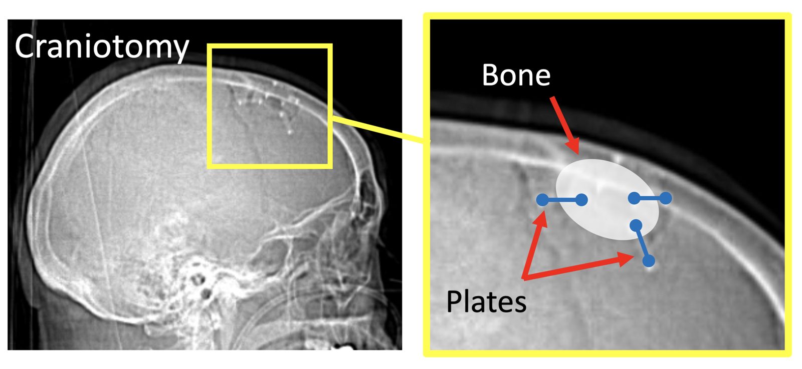
What kinds of brain lesions are treated with craniotomy?
Here are some example lesions.
My approach to craniotomy
My philosophy is to use the smallest possible craniotomy to safely achieve the goals of surgery. Smaller craniotomies are less invasive and easier to recover from.
I tend to use “keyhole” craniotomies, often about the size of a large coin, though the size depends on the location and nature of the lesion being treated. Ultimately, the driving factor is safety.
I use a variety of advanced technologies during craniotomies, including 3D navigation, microscopy, fluorescence, intraoperative ultrasound, and motor mapping. Awake craniotomies are reserved for cases in which a lesion is close to one of the speech centers of the brain and speech mapping is needed.
I shave only the minimum amount of hair needed, usually a short strip along the incision that is just wider than a Band-Aid. I do not shave the entire head.
What to expect from surgery
- For more detail about preparing for surgery and recovering afterward, please see my patient guide for craniotomy.
- Most craniotomies are performed with you asleep, under general anesthesia.
- The length of surgery varies with the location and nature of the lesion. Please discuss this with me at your visit.
- Most patients recover in the ICU the first night and stay in the hospital 1-2 nights.
Alternatives to craniotomy
Alternatives to craniotomy depend on the nature of the problem we are treating. Some small or slow-growing lesions may not need surgery at all and can be followed with surveillance MRIs. Lesions requiring treatment can sometimes be managed with radiation, such as Gamma Knife, with laser ablation, or with medications. I will go over the options that make sense for you at your visit.
Risks
The general risks of craniotomy include infection or an issue with wound healing, bleeding, spinal fluid leak, seizures, and new neurologic problems. These risks depend primarily on the location, size, and nature of the lesion being treated. Lesions that are removed can sometimes come back, depending on their underlying biological aggressiveness. I will review the pertinent risks of craniotomy specific to you before your surgery.