Laser Interstitial Thermal Therapy
-
Laser interstitial thermal therapy, also referred to as laser ablation, is a minimally invasive technique for precisely burning tissue in the body. It can be used in various organs, including the liver and prostate; I use it for certain disorders of the brain.
There are two main steps in a laser ablation procedure. In the first step, a small, temporary anchor is affixed to the skull. The anchor is inserted with millimeter precision, and it serves to aim the laser fiber toward a target in the brain. In the second step of the procedure, a laser fiber is inserted with the patient asleep in an MRI machine. The laser is then used to coagulate (burn) diseased brain tissue using the MRI to provide real-time feedback.
-
Laser ablation is a minimally invasive alternative to craniotomy that can be used for a variety of brain lesions. I use it most commonly for epilepsy. It is a flexible tool that can also be used for lesions such as cavernous malformations, certain brain tumors, and radiation necrosis. Laser ablation is best-suited for small lesions in the brain.
Below on the page is an image of a case I performed illustrating what a laser ablation lesion looks like on an MRI after the procedure.
-
Laser ablation is performed under general anesthesia. There is no need to shave more than a tiny patch of hair, and the incision is only a few millimeters long and can be closed with a single absorbable stitch. Patients generally stay one night in the hospital and can leave the following day. Headaches are common in the first few days after the procedure; these tend to respond to steroid medications, which I give to all patients.
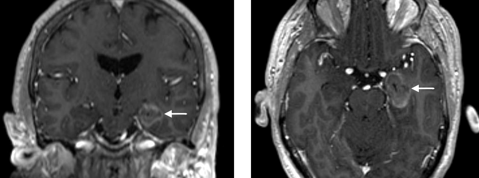
What a laser ablation lesion looks like on an MRI after the procedure

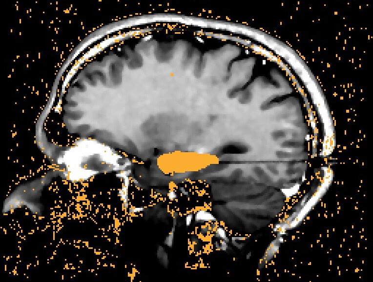
Example of how the MRI is used to monitor temperature change in the brain tissue that we are targeting in a laser ablation procedure. Images courtesy of Medtronic.

Photograph of the laser ablation system I use in my practice. Image courtesy of Medtronic.