Microvascular Decompression
Below is general information about microvascular decompression (MVD). I have a particular interest and specialized training in disorders treated with MVD, including trigeminal neuralgia, glossopharyngeal neuralgia, and hemifacial spasm.
I perform a high volume of MVDs, and I am passionate about the procedure. If you live near Denver and have a condition that might be treated with MVD, please consider meeting me in consultation to learn about the procedure and your options.
Click here to read the guide that I provide patients before surgery. Below are videos of microvascular decompressions that I have posted on YouTube.
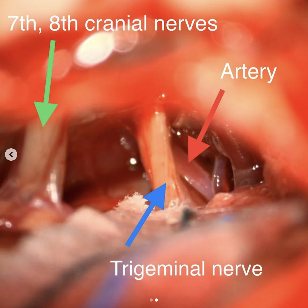
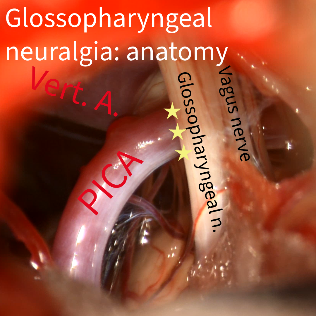
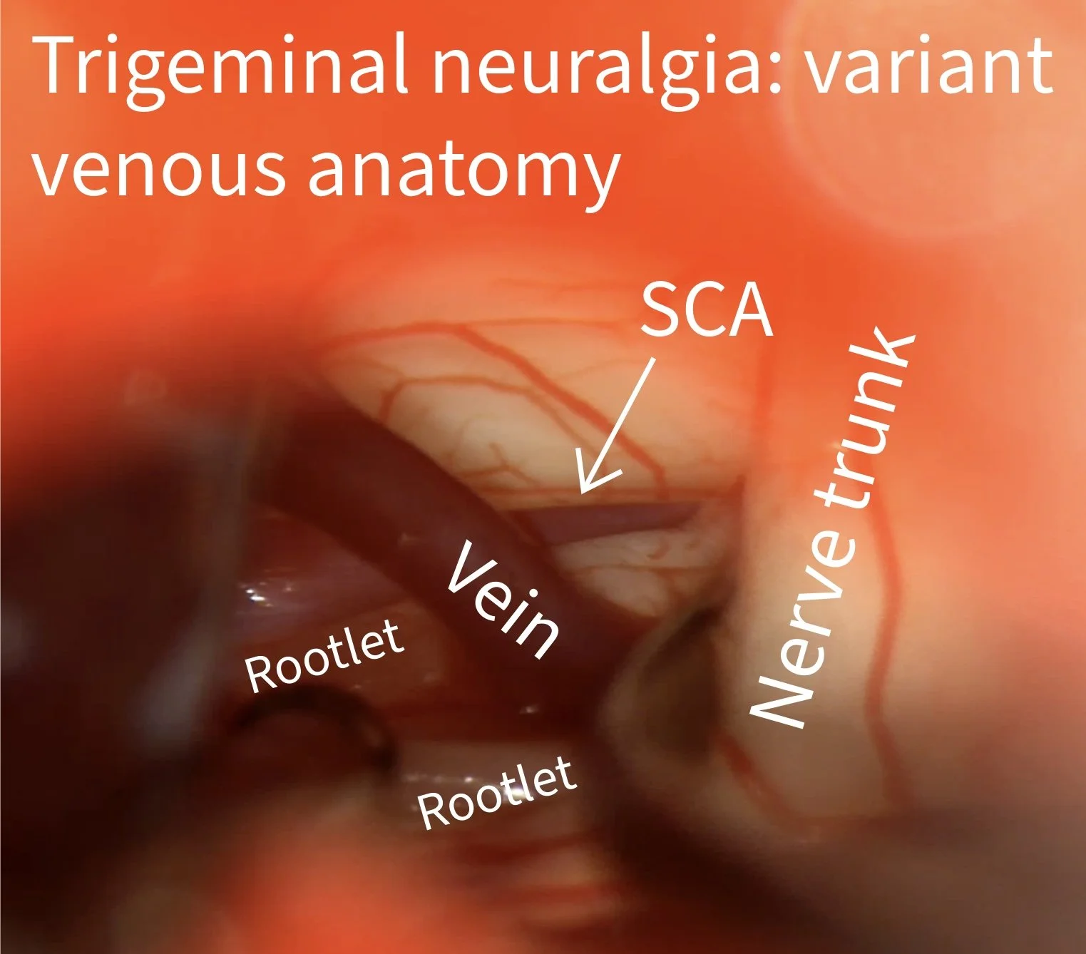
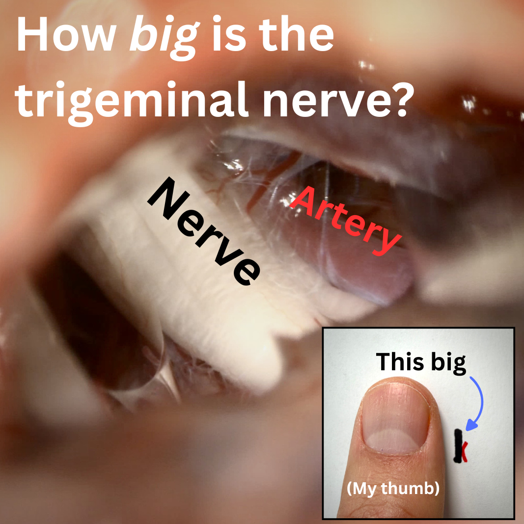
A microvascular decompression (MVD) is a type of craniotomy in which the nerves and blood vessels near the base of the brain are accessed using a small opening behind the ear. The goal of the operation is to identify and relieve compression of the nerves by a blood vessel. Commonly affected cranial nerves include the trigeminal nerve, the facial nerve, and the glossopharyngeal nerve.
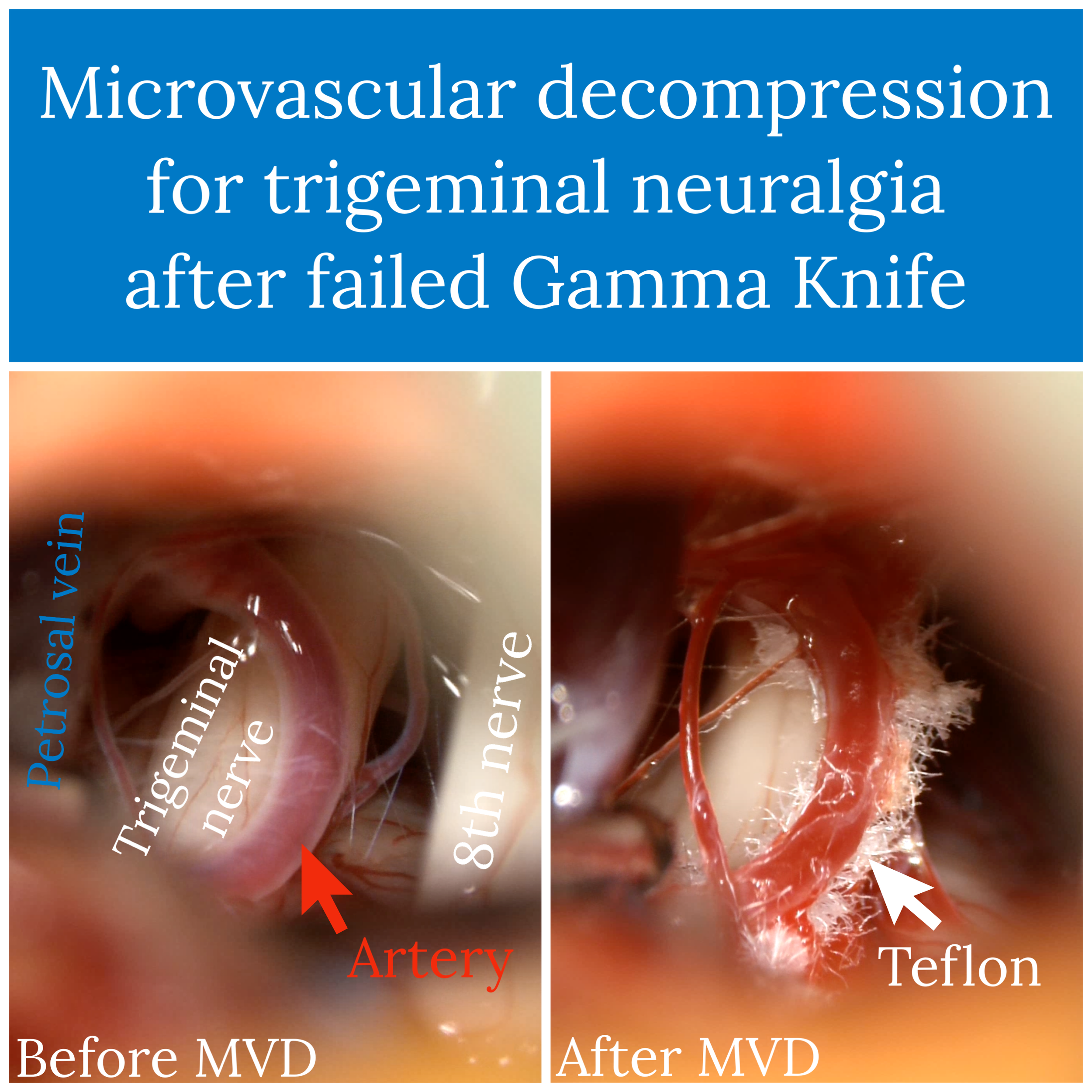
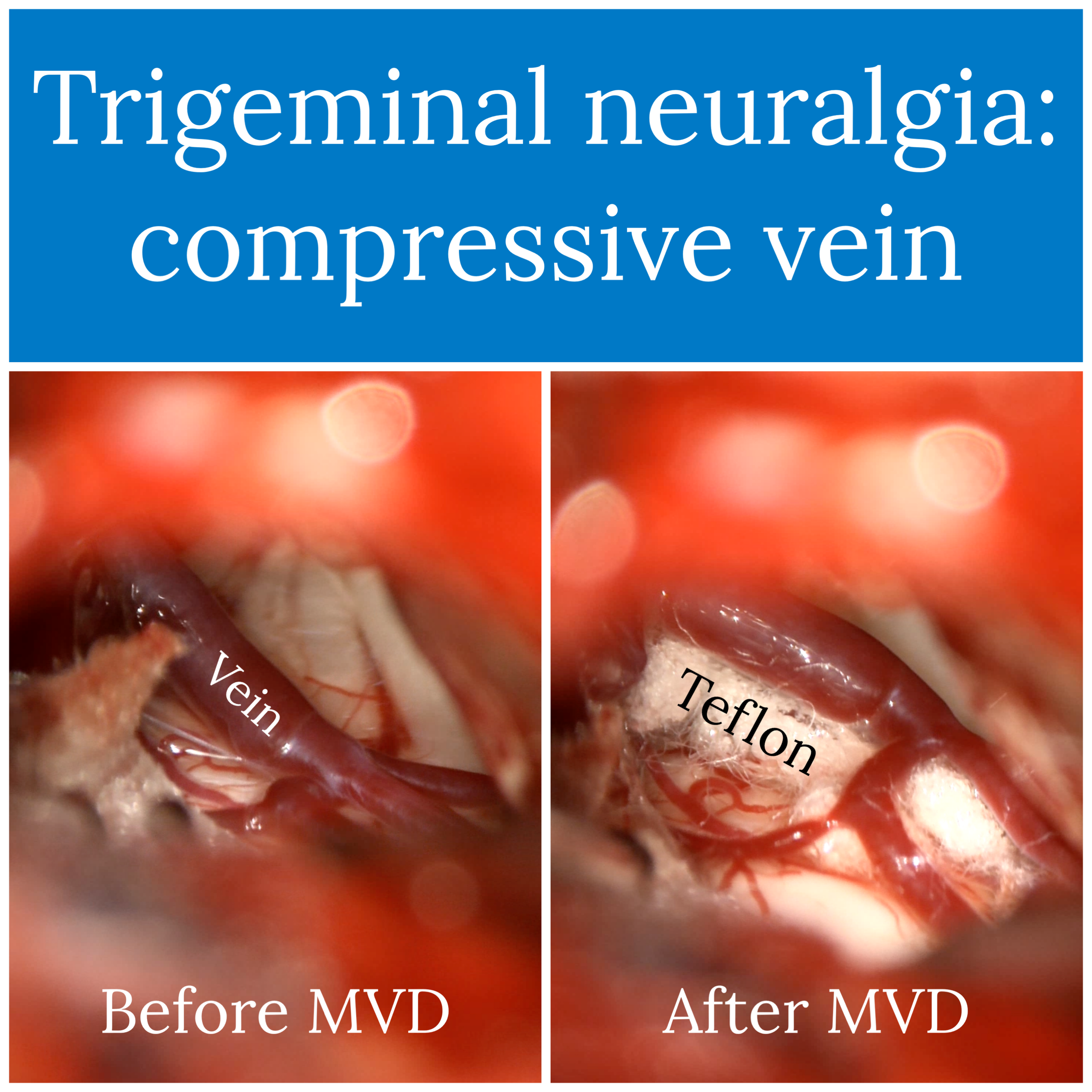
In an MVD, I drill an opening in the skull behind the ear that is about the size of a quarter. A microscope is used to identify the blood vessel and the nerve. They are dissected apart, and a piece of teflon padding is usually inserted between them (see photos, below). The hole in the bone is then repaired with bone cement.
MVD is used most commonly for trigeminal neuralgia, hemifacial spasm, and glossopharyngeal neuralgia, which are collectively referred to as neurovascular compression syndromes.
MVD has a high success rate for the disorders listed above. In trigeminal neuralgia, for example, MVD yields good or excellent pain relief in over 90% of patients and has the greatest durability of available surgical therapies. It is also the only first-line surgery for trigeminal neuralgia that does not involve damaging the trigeminal nerve. Success rates in hemifacial spasm are similar, with 80-90% of patients experiencing immediate spasm relief.
The recovery period after MVD involves a stay of 1-2 nights in the hospital. Some patients have a few hours of nausea after the procedure. Headache and incisional soreness are common; these tend to resolve over a few days. Most patients can expect to return to everyday activities around the home in the days after leaving the hospital. I ask patients to refrain from heavy lifting and vigorous exercise for 2-3 weeks. Resumption of work is guided by how you feel and varies depending on your age, overall health, and occupation.
If you live near Denver and have a disorder that is treated with MVD (trigeminal neuralgia, hemifacial spasm, or glossopharyngeal neuralgia), please consider meeting me in consultation to learn more about treatment options.