
Endoscopic Third Ventriculostomy (ETV)
The following is for general educational purposes only. I no longer accept referrals for hydrocephalus, VP shunts, or related disorders.
Endoscopic third ventriculostomy (abbreviated ETV) is a procedure for treating hydrocephalus.
Hydrocephalus is a disorder in which spinal fluid accumulates in the brain under pressure. Sometimes this is due to a blockage in the outflow of spinal fluid. Symptoms may include headaches, visual changes, nausea, and lethargy. ETV involves creating an alternate route for spinal fluid to drain internally around the site of a blockage. The procedure is an alternative to a ventriculoperitoneal shunt.
In an ETV, a tiny camera is inserted into the fluid-filled cavities in the brain (the ventricles), and an instrument is used to punch a hole in a thin membrane along the floor of the third ventricle, creating an alternate route for spinal fluid to flow out of the brain and into the spinal canal.
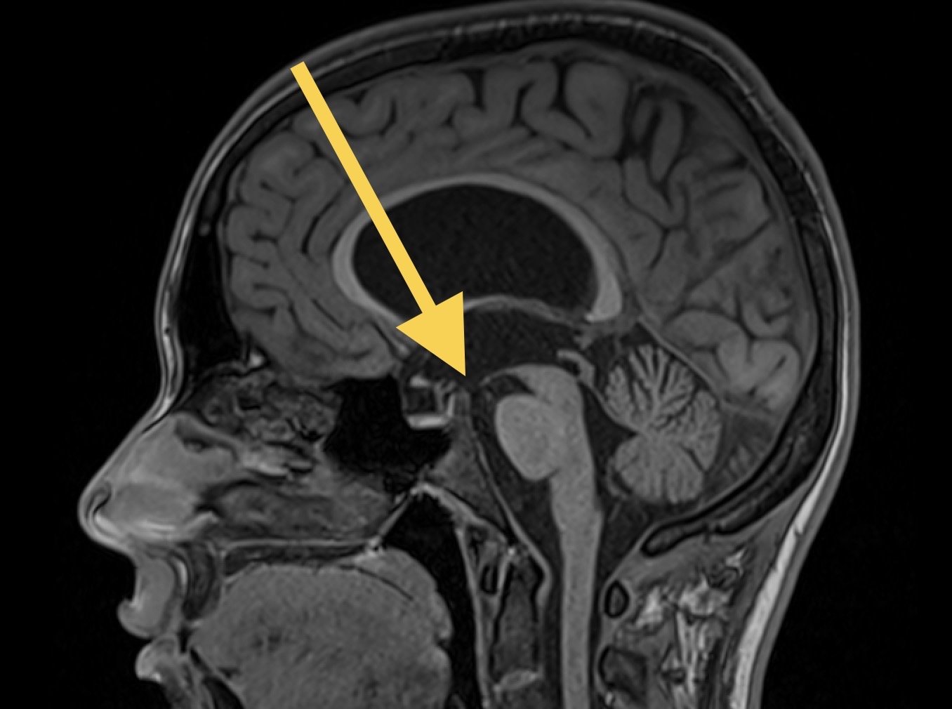
Below is an MRI from a patient with hydrocephalus as well as a view from the endoscope of the floor of the third ventricle. The yellow arrows indicate the path of the camera and the membrane where the hole is punched. On the photograph, the * indicates the approximate location the hole is punched in the floor of the third ventricle. Also visible is the basilar artery (B), the clivus (C), the infundibular recess (I; origin of the pituitary gland), and the mamillary bodies (M).


View during an endoscopic third ventriculostomy showing relevant anatomy including the mammillary bodies (M), basilar artery (B), clivus (C), infundibular recess (I), and the floor of the third ventricle (*).

View during an endoscopic third ventriculostomy showing relevant anatomy including the mammillary bodies (MB), basilar artery, infundibular recess (IR), optic chiasm, and the floor of the third ventricle where a stoma was created.
A major advantage of ETV over a ventriculoperitoneal shunt is it does not require a permanent implant. Shunts are effective, but they can sometimes clog or malfunction over time and need to be replaced.
Importantly, not all patients with hydrocephalus are candidates for an ETV. A patient’s brain anatomy, history of previous surgery, symptoms, and the reason for their hydrocephalus all factor into my evaluation.